Thank you Colombia

Full house at CVDC

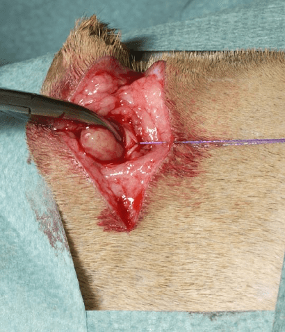
Thank you Colombia for a warm welcome and an excellently organised CVDC conference. More than 5000 vets (plus 3000 online) were there and at least 1750 vets visited my lectures! Truly amazing. Question of the day: what is a good flap to close wounds in the inhumanly area. Answer: if the skinfolds are intact, than I would always use a skinfold rotational flap. This is such a versatile technique.

It was first described by Hunt GB, Tisdall PL, Liptak JM, Beck JA, Swinney GR, Malik R. Skin-fold advancement flaps for closing large proximal limb and trunk defects in dogs and cats. Vet Surg. 2001 Sep-Oct;30(5):440-8.

In their summary they describe six dogs and 2 cats that underwent reconstruction of soft-tissue wounds resulting from traumatic, neoplastic, or infectious lesions. Skin-fold flaps were created by division of the medial and lateral attachment to the proximal limb or the dorsal and ventral attachment to the trunk, enabling closure of adjacent defects on the trunk or proximal limb, respectively.

Their conclusion was The skin-fold advancement flap is a versatile technique that lends itself to use in a variety of locations, depending on which attachments are divided. The clinical results are comparable with those reported for axial pattern and subdermal plexus flaps.

Coffee country

Ivanov in front if the Tio Conejo coffee stand

If you are in Perreira you need to drink coffee. Pereira is the capital city of the Risaralda Department, located in the coffee-growing region of the country. It is part of the “Coffee Axis” along with the cities of Armenia and Manizales.

Pereira is known for its pleasant climate, surrounded by lush green landscapes, and it serves as a hub for commerce, agriculture, and tourism. If you are indeed referring to Pereira, it is a beautiful city with a rich cultural heritage, making it an interesting destination for travelers.

I really love it here and the coffee of course. My friend Ivanov Castellanos has his own coffee plantation and I had many a cup! I also gave 2 lectures at CVDC about mammary tumors!

Mammary tumors in cats are a relatively common occurrence, particularly in older female cats that haven’t been spayed. These tumors are abnormal growths that develop in the mammary glands, which are the milk-producing glands found in the breast tissue.

Here are some key points about mammary tumors in cats:

  1. Prevalence: Unspayed female cats are at a higher risk of developing mammary tumors. The risk increases with age, and most tumors occur in cats over 10 years old.
  2. Malignancy: Approximately 85-90% of feline mammary tumors are malignant (cancerous). Therefore, it’s essential to identify and treat them early to increase the chances of successful treatment.
  3. Symptoms: Mammary tumors may present as firm, often painless masses in the mammary gland area. They can be single or multiple and are usually found under the skin.
  4. Diagnosis: If you notice any abnormal masses or changes in the mammary gland area of your cat, it’s essential to seek veterinary attention promptly. Your veterinarian will perform a physical examination, potentially take a fine-needle aspiration or biopsy for further evaluation.
  5. Treatment: Treatment options for mammary tumors in cats may include surgical removal of the tumor, possible removal of the entire mammary gland chain, or other treatments like radiation or chemotherapy in more advanced cases.
  6. Prognosis: The prognosis for mammary tumors in cats depends on various factors, such as tumor size, type, and stage at the time of diagnosis. Early detection and prompt treatment generally lead to better outcomes.

Having a great time in Bogota

Yesterday I had the honor to speak for the students of La Salle University Norte in Bigot, Colombia. We talked about how to do a correct biopsy and how important a true diagnosis of the type of cancer is for your treatment planning

We had so many excellent questions: do you worry about seeding tumor during Fine needle aspirates? My answer: there is always a risk but the risk is super small and the advantage of FNAB weighs way up against the chance of spreading the tumor!

Fine needle aspirates (FNA) play a crucial role in tumor detection in pets. Veterinarians use FNA as a minimally invasive diagnostic tool to collect cell samples from suspicious masses or tumors in animals. This procedure involves inserting a thin needle into the mass, extracting cells, and then examining them under a microscope. FNA helps determine whether the mass is benign or malignant, aiding in early diagnosis and guiding appropriate treatment options for the pet’s condition.

New podcast is out!

Yippee, our new podcast is out after 2 years of silence. Our first article of the year will discuss the surgical description and outcome of ultrasound-guided minimally invasive parathyroidectomy in 50 dogs with primary hyperparathyroidism. We will have a short interview with Alasdair Hotson Moore and some surgery news. Download at any of the podcast platforms.

Polyps in the ear

Nasopharyngeal polyps, also called otopharyngeal or inflammatory polyps, are benign pedunculated growths of uncertain origin but thought to arise as a result of chronic inflammation. Polyps have been associated with rhinitis and otitis resulting from various bacterial and viral agents; a congenital origin has been suggested as well. They may originate from the mucosal lining of the middle ear, auditory tube, and nasopharynx, all of which are of similar histologic origin. Otopharyngeal polyps occur in cats of any age, although most animals are less than 2-years old. Polyps in the external or middle ear mimic signs of otitis externa, otitis media or otitis interna.

Otoscopy after flushing may reveal a visible pink or grey smooth, spherical mass occluding the canal. Cytologic or histologic examination of biopsies will reveal the nature of the tissue when the diagnosis is not straightforward. Some surgeons perform a ventral bulla osteotomy in all cases, but this is rarely indicated because recurrence is uncommon with simple traction-avulsion after an incision in the vertical ear canal.

Vertical ear canal approach of an inflammatory polyp

For more information, check out our basic surgery book, Chapter 14:

https://online.flippingbook.com/view/215593981/