
I’m not entirely sure how the topic came up during my half-day whirlwind tour of Jakarta (thank you, TripAdvisor!), but there I was—sweating through my eltaMD sunscreen—when my delightful guide Stella was trying to explain a lively celebration happening in a port neighborhood. After much thoughtful vocabulary searching, she landed on a rather unexpected word: circumcision. Yup, you read that right. I wonder how I got that description wrong? Turns out, it’s common here to throw a full-blown party for the occasion. Welcome to Jakarta!
With a population of over 12 million, and a large Muslim majority, I suppose I shouldn’t have been all that surprised. Still, not exactly on the top of my sightseeing bingo card.
Our tour also coincided with Jakarta’s birthday—June 14, a date that traces back to 1527. That means a 500-year anniversary in 2027, and I’m already mentally RSVPing. There are personal ties too: both my grandparents once lived here, and my mom was born in Batavia, as Jakarta was known in its Dutch colonial days. The name Jakarta itself means “City of Victory,” which sounds just dramatic enough for a place like this.
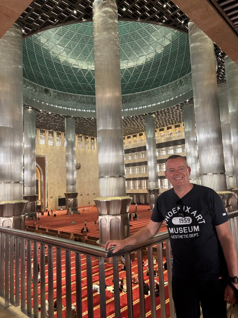
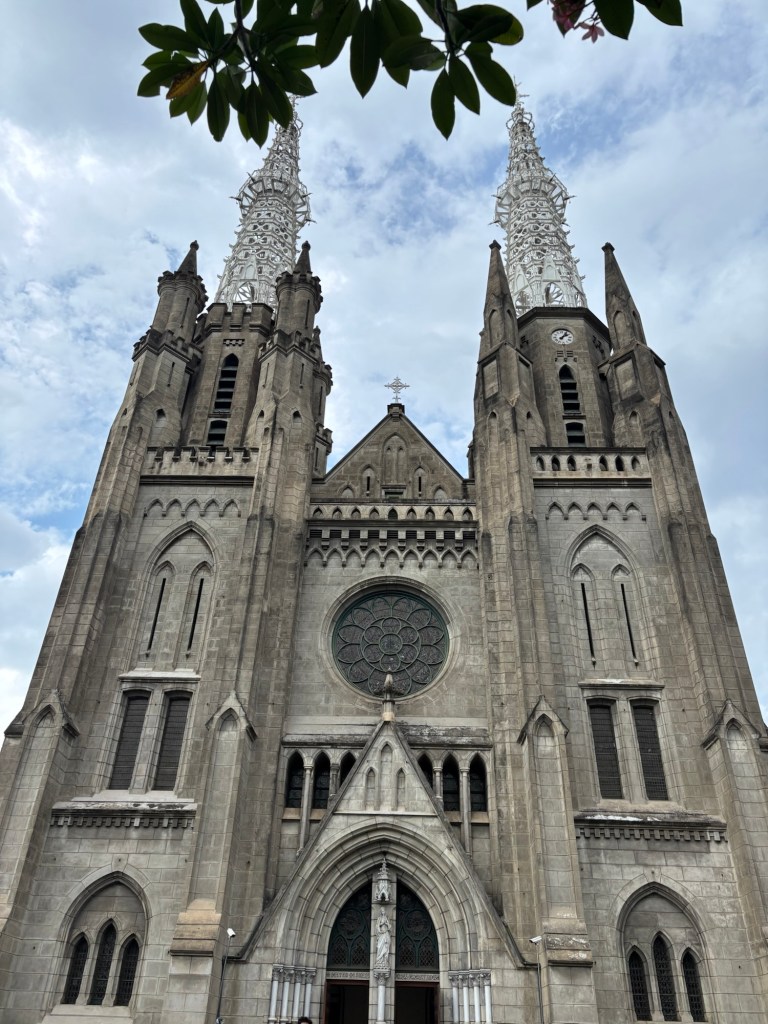
First stop: the Istiqlal Mosque—“Istiqlal” means independence, and the building is as symbolic as it is impressive. Fun fact: it was designed by a Christian architect. The dome spans 45 meters, representing 1945, the year of Indonesia’s independence. Right across the street? A Catholic cathedral, proudly parked there as a shining example that yes, different faiths can live peacefully side-by-side—sometimes just across the road.
The cathedral itself is like a hybrid: Protestant restraint meets Catholic bling. A little wooden cross, a little gold, something for everyone.
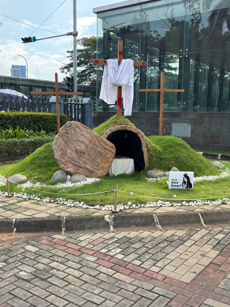
Outside, there’s a small hill with three crosses and a stone dramatically rolled to the side—which, let’s be honest, probably moonlighted as part of a Pesach-themed stage set at some point.
As we zipped through the city (and I mean zipped—Jakarta’s traffic is basically an extreme sport), I couldn’t help but marvel at the moped madness. Especially the green-jacketed drivers, who are basically Indonesia’s version of Uber: bookable via apps like Gojek or Grab, or just flag one down and hang on tight.
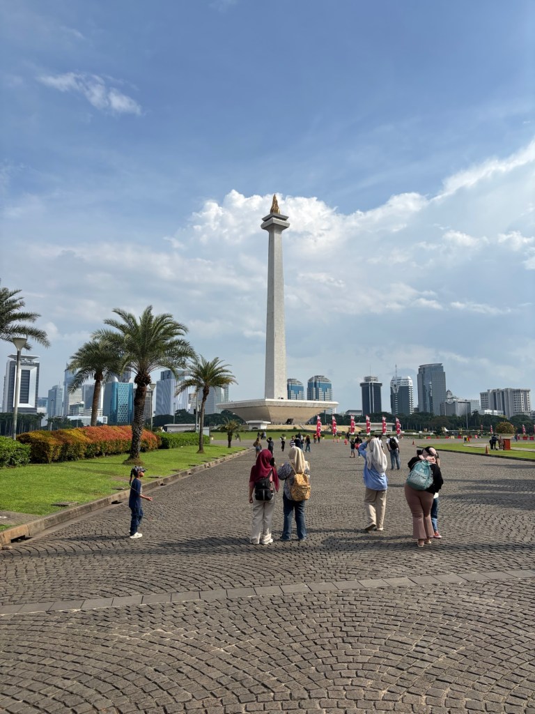
Next up: the National Monument, or Monas, a giant spire with a golden flame on top. You access the base via an underground tunnel, which is wonderful, because walking above ground at noon here feels like being sautéed in your own shoes. Inside, the story of Indonesia’s fight for independence is told in little dioramas. Let’s just say… the Dutch aren’t exactly the heroes of this tale. But don’t worry, Stella assures me: the Japanese occupation was apparently even worse. Yay?
And although the Dutch didn’t leave the best political legacy, they certainly left a lot of vocabulary. Stella rattled off a list:
• Gordijn = curtain
• Waskom = wash basin
• Benzine = gasoline
• Kantoor = office
• Koffiehuis = coffee house
You’re welcome, Bahasa Indonesia.
Jakarta’s streets are a universe of their own. Unofficial traffic guides wander between the cars, helping you squeeze through the chaos—for a small fee. Skip the tip and, according to Stella, your car may get a “mystery scratch.” Noted.
Of course, not everything sparkles. Poverty is very present, and a lot of people live below the poverty line. And while the Dutch did attempt to recreate their beloved canals here, in Jakarta’s sweltering humidity those charming waterways turned into stagnant, mosquito-infested puddles of doom.
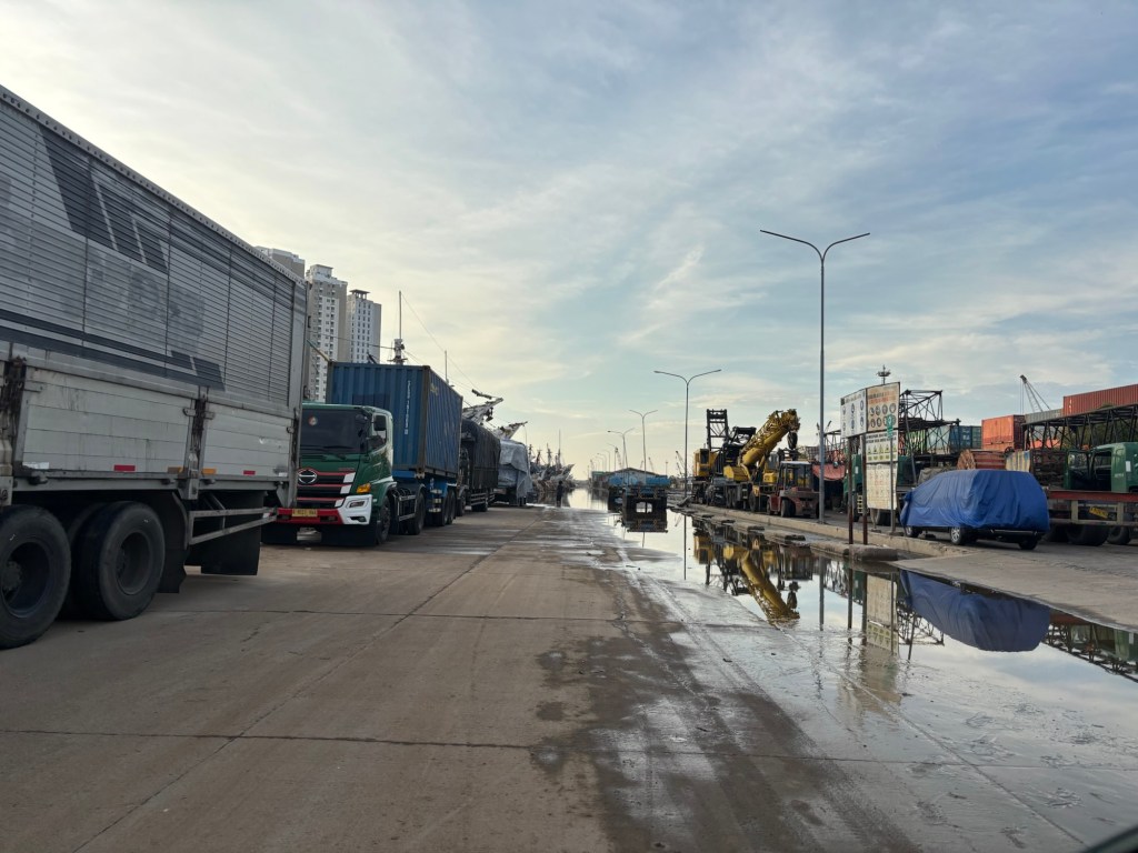
To make matters more dramatic, Jakarta is sinking—fast. Thanks to relentless groundwater pumping and unchecked construction, the city is literally dropping by as much as 10 cm a year, making it one of the fastest-sinking cities on the planet. When we visited the harbor, part of it was already underwater. Gigantic sea walls now line the coast in an attempt to keep the ocean from claiming more real estate.
On our way back, Stella gave me a final warning: “Don’t go out on the main street after 9 p.m.—too dangerous,” she said. I nodded gravely, all while knowing full well that jet lag has me drooling on my pillow by 8:45 anyway.